- Chemical equation
- Question And Answers
- Chemical equation
- Law of conservation of mass
- Laws of constant proportion
- Dalton’s atomic theory
- Atoms and molecules
- Symbols of elements
- Atomicity
- Valency
- What is an Ion ?
- Atomic mass
- Molecules of compounds
- Molecular mass , Formula unit mass
- Mole
- Molar mass
- Types of Chemical Reactions
- Decompostion Reaction
- Displacement reaction
- Double displacement reaction
- Oxidation and Reduction
- Effects of oxidation reactions in daily life
- 7.Atoms, Molecules And Chemical Reactions
- Atoms, Molecules, and chemical reactions
- Classification of Elements
- Dobereiner’s law of Triads
- Limitations
- Mendeleeff’s Periodic Table
- Salient features and achievements of the Mendeleeff’s periodic table
- Limitations of Mendeleeff’s periodic table
- Modern Periodic Table
- Groups,
- Periods
- Metals and Non metals
- Atomic radius
- Periodic properties of the elements in the modern table
- Ionization Energy
- Electronegativity
- Metallic and Non-Metallic Properties
- Short answer Questions
- Question And Answers
- Synopsis
- Classification of elements - The Perodic Table
- Nutrition
- Mechanism of Photosynthesis
- Light independent reaction (Biosynthetic phase)
- Heterotrophic nutrition and nutrition in Human Beings
- Health aspects of the elementary canal
- Coordination in life processes
- Peristaltic Movement in Esophagus
- Taste connected with tongue and palate
- Villi
- Respiration -The energy Producing system
- Epiglottis and passage of air
- Gaseous Exchange
- Respiration versus combustion
- Transportation-The circulatory system
- The blood vessels and The cardiac cycle
- Blood pressure and Materials transport in the plants
- The wastage disposing system
- Mechanisms of urine formation
- Other Pathways Of Excretion
- Excretion release in substance of plants
- Short Answer Questions
- Long answer Questions
- Synopsis
- Reproduction - The generating system
- Reproduction in a placental mammal - Man
- Cell cycle
- Reproduction In Organisms
- Parthenogenesis
- Vegetative Propagation
- Male Reproductive System
- Female reproductive system
- STRUCTURE OF A SPERM
- Menstrual Cycle
- Extra Embryonic Membrane
- Sexual reproduction in flowering plants
- Synopsis
- Long Answer Questions
- Short Answer Questions
- Quizz
- Important Quiz Questions
- Introduction
- Lewis Electron Dot Symbols
- Ionic Compounds: Electrons Transferred
- Formation of Ionic Compounds
- The arrangement of ions in ionic compounds
- Covalent bond
- VSEPR theory
- Valence bond theory
-
Formation of O_2, N_2, CH_4Molecules
- Hybridisation
- Properties of ionic and covalent compounds
-
Formation of BF_3,NH_3, water molecule
- Synopsis
- Question And Answers
- Chemical Bonding
Transportation-The circulatory system
All the living organisms need nutrients, gases, liquids etc., for growth and maintenance of the body.
All the organisms would need to send these materials to all parts of their body whether they are unicellular organisms or multicellular.
- In unicellular organisms these may not have to be transported to longer distances while in multicellular forms have to be sent substances to long distances as far as say over 100 feet for the tallest plant on earth.
- In lower organisms like amoeba, hydra etc., all the materials are transported through a simple process like diffusion, osmosis etc.,
- In higher animals with trillions of cells in their body adopt the method of diffusion and osmosis only for the bulk movement of materials, would takes years.
- To avoid delay a separate system is needed to carry the materials much faster and more efficiently.
This specialized system that is developed by organisms is called ‘the circulatory system’.
In the year 1816, Rene Laennec discovered the Stethoscope. Before the discovery of stethoscope doctors used to hear the heartbeat by keeping an ear on the chest of the patient. Laennac found that paper tube helps to hear
the heart beat perfectly. Then he used a bamboo instead of paper tube to
hear heartbeat. Laennac called it the stethoscope
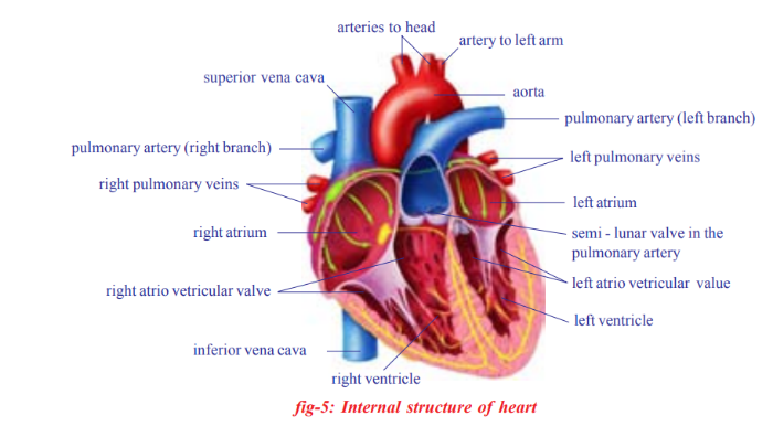
Internal structure of the heart
• Keep the heart in the tray in such a way that a large arch-like tube faces upwards. This is the ventral side.
• Now take a sharp blade or scalpel and open the heart in such a way that the chambers are exposed

- The heart is a pear shaped structure, triangle in outline, wider at the anterior end and narrower at the posterior end.
- The heart is covered by two layers of membranes. The membranes are called pericardial membranes. The space between these two layers is filled with pericardial fluid, which protects the heart from shocks.
- The heart is divided into four parts by grooves.
Two upper parts are called atria (auricles), and the lower ones are called
ventricles. - The left atrium and ventricle are smaller when compared to that of
right atrium and ventricle. - The blood vessels found in the walls of the heart are coronary vessels which supply blood to the muscles of the heart.
- The walls of the ventricles are relatively thicker than atrial walls.
- the heart has four chambers in it. On the left side two chambers are present, one is anterior and the other is the posterior. On the right side also two chambers present, one upper (anterior), and one lower (posterior)
- The rigid vessels are called arteries which originate from the heart and supply blood to various organs in the body. The larger artery is the aorta.
- The relatively smaller one is pulmonary artery which carries blood from the heart to the lungs.
- The less rigid vessels are the veins, which bring blood from all body parts to the heart.
- The vein which is at the anterior end of the right side of the heart is superior venacava (precaval vein), which collects blood from anterior parts of the body.
- The vein which is coming from posterior part of the heart is inferior venacava (postcaval vein), collecting blood from posterior part of the body.
- The two atria and the two ventricles are separated from each other by muscular partitions called septa. The openings between atria and ventricles are guarded by valves.
- In the right atrium we can observe the openings of superior and inferior venacava. In the left atrium, we can observe the openings of pulmonary veins, that bring blood from lungs.
- From the upper part of the left ventricle, a thick blood vessel called aorta arises. It supplies oxygenated blood to the body parts.
- From the upper part of the right ventricle-pulmonary artery arises that supplies deoxygenated blood to the lungs. After careful examination, we can observe valves in the pulmonary artery and aorta as well.


0 Doubts's